d o t o r e g . c o m
Right Arm - Left Leg Lead Reversal
 May give the impression of old INFERIOR myocardial infarction at first glance.
On the other hand, it may mask the ECG signs old INFERIOR myocardial infarction,
even if the patient really had one.
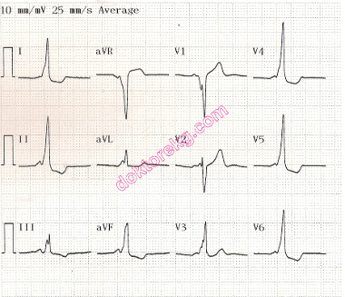
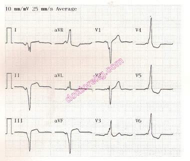
ECGs 1a (left) and 1b (right). The compact ECGs above belong to a 27 years-old woman with WPW syndrome.
ECG 1a was recorded with correctly positioned electrodes. ECG 1b was recorded with reverse connection of the right arm and
left leg electrodes.
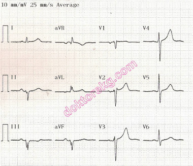
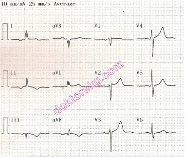
ECGs 2a (left) and 2b (right). The compact ECGs above belong to a 51 years-old man with who had experienced
old inferior myocardial infarction (due to Circumflex coronary artery obstruction) 3 years ago.
ECG 2a was recorded with correctly positioned electrodes. ECG 2b was recorded with reverse connection of the right arm and
left leg electrodes.
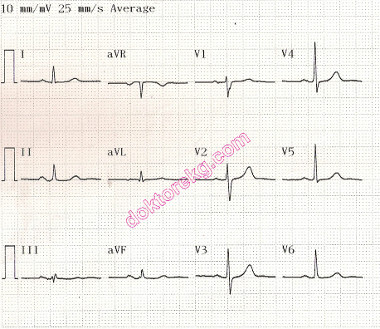
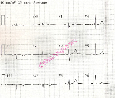
ECGs 3a (left) and 3b (right). The compact ECGs above belong to a 65 years-old man with systemic arterial hypertension,
coronary artery disease and chronic obstructive pulmonary disease (COPD). A stent had been previously implanted to his
right coronary artery (RCA), however he had never had an acute myocardial infarction before.
ECG 3a was recorded with correctly positioned electrodes. ECG 3b was recorded with reverse connection of the right arm
and left leg electrodes. Right arm-left leg lead reversal gives the impression of old inferior myocardial infarction.
May give the impression of old INFERIOR myocardial infarction at first glance.
On the other hand, it may mask the ECG signs old INFERIOR myocardial infarction,
even if the patient really had one.
ECGs 1a (left) and 1b (right). The compact ECGs above belong to a 27 years-old woman with WPW syndrome.
ECG 1a was recorded with correctly positioned electrodes. ECG 1b was recorded with reverse connection of the right arm and
left leg electrodes.
ECGs 2a (left) and 2b (right). The compact ECGs above belong to a 51 years-old man with who had experienced
old inferior myocardial infarction (due to Circumflex coronary artery obstruction) 3 years ago.
ECG 2a was recorded with correctly positioned electrodes. ECG 2b was recorded with reverse connection of the right arm and
left leg electrodes.
ECGs 3a (left) and 3b (right). The compact ECGs above belong to a 65 years-old man with systemic arterial hypertension,
coronary artery disease and chronic obstructive pulmonary disease (COPD). A stent had been previously implanted to his
right coronary artery (RCA), however he had never had an acute myocardial infarction before.
ECG 3a was recorded with correctly positioned electrodes. ECG 3b was recorded with reverse connection of the right arm
and left leg electrodes. Right arm-left leg lead reversal gives the impression of old inferior myocardial infarction.