d o t o r e g . c o m
Dr. Mete ALPASLAN
Samples of Abnormal Treadmill Exercise Test
Pages 1 - 2 - 3 - 4 - 5 - 6
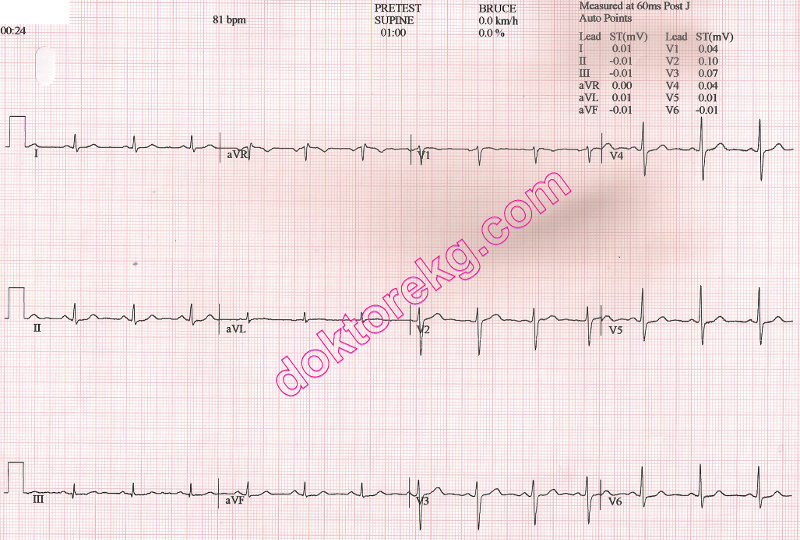
ECG 11a. The above ECG is from a 65 years-old man.
It was recorded just before the onset of his treadmill exercise test.
He was complaining of chest oppression (unrelated to effort) for the last 3 days.
Click here for a more detailed ECG

ECG 11b. The above ECG belongs to the same patient.
It was recorded at the 3rd minute of his treadmill exercise test.
Leads V4 to V6 show ST segment depression.
Click here for a more detailed ECG

ECG 11c. The above ECG belongs to the same patient.
It was recorded at the 6th minute of his treadmill exercise test.
Leads V4 to V6 show ST segment depression.
Click here for a more detailed ECG

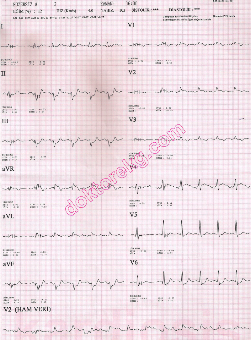
ECG 11d. The above ECG belongs to the same patient.
It was recorded at the 9th minute of his treadmill exercise test.
Lead V6 shows ST depression clearly. Most of the other leads show baseline drift.
Click here for a more detailed ECG
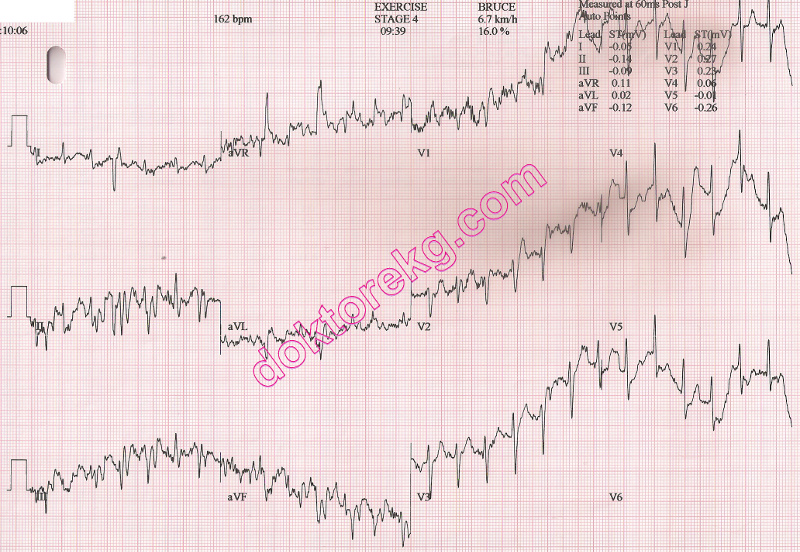
ECG 11e. The above ECG belongs to the same patient.
It was recorded 9 minutes and 39 seconds after the onset of his treadmill exercise test.
This is his peak exercise. The treadmill was stopped at this time.
Extensive baseline drift prevents commenting.
Click here for a more detailed ECG
ECG 11f. The above ECG belongs to the same patient.
It was recorded 3 minutes after the termination of his test (recovery phase).
ST segment depression is seen in leads I, II, V5 and V6.
Click here for a more detailed ECG

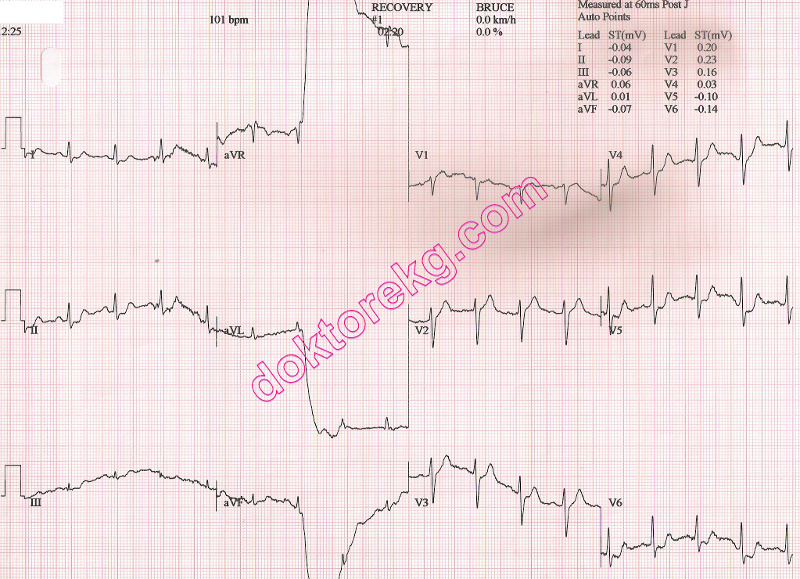
ECG 11g. The above ECG belongs to the same patient.
It was recorded 4 minutes after the termination of his test (recovery phase).
ST segment depression is seen in leads I, II, III, aVF, V5 and V6.
Lead aVR shows ST segment elevation.
Click here for a more detailed ECG
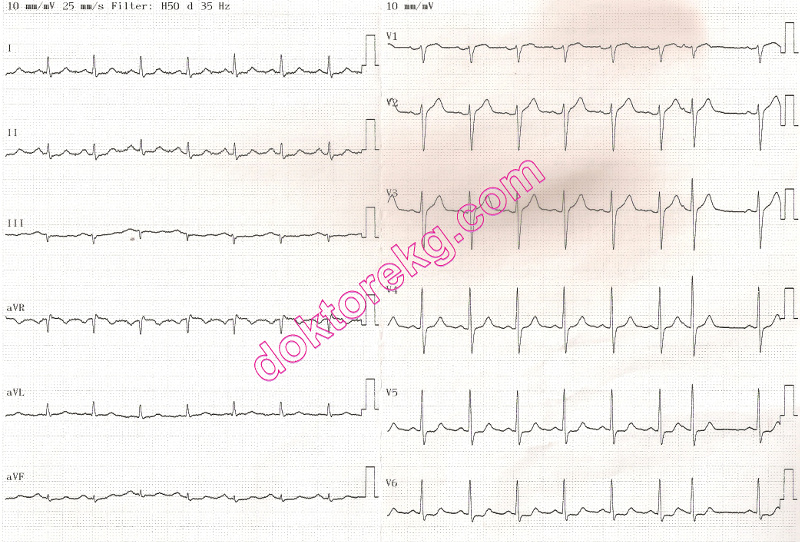
ECG 11h. The ECG above belongs to the same patient. It was recorded 30m minutes after termination of
the treamil exercise test. He was experiencing chest oppression at this time.
Leads I, aVF, V5 and V6 show ST segment depression while lead aVR shows ST segment elevation.
He underwent coronary angiography 30 minutes after recording of the above ECG.
Hemodynamically significant stenoses of the LAD artery and its Diagonal branch were observed.
Click here for a more detailed ECG

ECG 12a. The resting ECG above belongs to a 62 years-old man with the complaint of effort angina.
Sinus bradycardia with a heart rate of 44/minute is seen.
Click here for a more detailed ECG
ECG 12b. The ECG above belongs to the same man. It was recorded at the sixth minute of his treadmill exercise test.
Leads II, III, aVF, V5 and V6 show upsloping type ST segment depression.
Leads V1 and V2 show ST segment elevation.
Click here for a more detailed ECG
Page 1 - Page 2 - Page 3 - Page 4 - Page 5 - Page 6
Pages 1 - 2 - 3 - 4 - 5 - 6
ECG 11a. The above ECG is from a 65 years-old man.
It was recorded just before the onset of his treadmill exercise test.
He was complaining of chest oppression (unrelated to effort) for the last 3 days.
Click here for a more detailed ECG
ECG 11b. The above ECG belongs to the same patient.
It was recorded at the 3rd minute of his treadmill exercise test.
Leads V4 to V6 show ST segment depression.
Click here for a more detailed ECG
ECG 11c. The above ECG belongs to the same patient.
It was recorded at the 6th minute of his treadmill exercise test.
Leads V4 to V6 show ST segment depression.
Click here for a more detailed ECG
ECG 11d. The above ECG belongs to the same patient.
It was recorded at the 9th minute of his treadmill exercise test.
Lead V6 shows ST depression clearly. Most of the other leads show baseline drift.
Click here for a more detailed ECG
ECG 11e. The above ECG belongs to the same patient.
It was recorded 9 minutes and 39 seconds after the onset of his treadmill exercise test.
This is his peak exercise. The treadmill was stopped at this time.
Extensive baseline drift prevents commenting.
Click here for a more detailed ECG
ECG 11f. The above ECG belongs to the same patient.
It was recorded 3 minutes after the termination of his test (recovery phase).
ST segment depression is seen in leads I, II, V5 and V6.
Click here for a more detailed ECG
ECG 11g. The above ECG belongs to the same patient.
It was recorded 4 minutes after the termination of his test (recovery phase).
ST segment depression is seen in leads I, II, III, aVF, V5 and V6.
Lead aVR shows ST segment elevation.
Click here for a more detailed ECG
ECG 11h. The ECG above belongs to the same patient. It was recorded 30m minutes after termination of
the treamil exercise test. He was experiencing chest oppression at this time.
Leads I, aVF, V5 and V6 show ST segment depression while lead aVR shows ST segment elevation.
He underwent coronary angiography 30 minutes after recording of the above ECG.
Hemodynamically significant stenoses of the LAD artery and its Diagonal branch were observed.
Click here for a more detailed ECG
ECG 12a. The resting ECG above belongs to a 62 years-old man with the complaint of effort angina.
Sinus bradycardia with a heart rate of 44/minute is seen.
Click here for a more detailed ECG
ECG 12b. The ECG above belongs to the same man. It was recorded at the sixth minute of his treadmill exercise test.
Leads II, III, aVF, V5 and V6 show upsloping type ST segment depression.
Leads V1 and V2 show ST segment elevation.
Click here for a more detailed ECG
Page 1 - Page 2 - Page 3 - Page 4 - Page 5 - Page 6